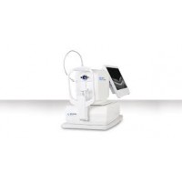
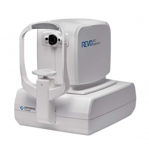
Revo
CT made simple as never before. All it must be done is to position the patient and press the START button to acquire examinations of both eyes. The device will make examination independently.
Small system footprint, various operator and patient positions allow to install SOCT Copernicus REVO even in the smallest examination room. Variety of review and analysis tool give the operator a choice of using it as a screening or as an advanced diagnostic device.
The noise reduction technology provides the finest details proven to be important for early disease detection.
RETINA
Single 3D Retina examination is enough to perform both Retina and Glaucoma analysis based on retinal scans. Software automatically recognize 8 retina layers. Thus allowing a more precise diagnosis and mapping any changes in the patient’s retina condition.


GLACUOMA
Comprehensive glaucoma analysis tools for Quantification of Optic Nerve Head, Retina Nerve Fiber Layer, DDLS, Ganglion layer and Asymmetry


ANTERIOR
For standard examinations no additional lens is required.
Additional adapter provided with the device allows to make wide scans of anterior segment.

FOLLOW UP
High density of standard 3D scan allows to precisely track the disease progress.
Operator can analyze changes is morphology, quantified progression maps or evaluate the progression trends.


NETWORKING
A proficient networking solution increases productivity and an enhanced patient experience. It allows you to view and manipulate multiple examinations from review stations in your practice. Effortlessly helping to facilitate patient education by allowing you to interactively show examination results to patients. Every practice will have differing requirements which we can provide by tailoring a bespoke service. There is no additional charge for the server module.
TECHNICAL DATA
| Technology | Spectral Domain OCT |
| Light Source | SLED, Wavelength 840nm |
| Bandwidth | 50 nm half bandwidth |
| Scanning speed | 60,000 A-scan per second - 110,000 A-Scan per second. |
| Axial resolution | 5 µm in tissue |
| Transverse Resolution | 12 µm, typical 18 µm |
| Overall scan depth | 2.4 mm |
| Scan range | 3 to 12 mm |
| Scan types | 3D, Radial, B-scan, Raster, Cross |
| Fundus image | Live Fundus Reconstruction |
| Alignment method | Fully automatic, Semi-automatic |
| Retina analysis | Retina thickness, Inner retinal thickness, Outer retinal thickness, RNFL+GCL+IPL thickness, GCL+IPL thickness, RNFL thickness, RPE deformation, IS/OS thickness |
| Glaucoma analysis | RNFL, ONH morphology, DDLS, Ganglion analysis as RNFL+GCL+IP and GCL+IPL, OU and Hemisphere asymmetry |
| Anterior | Pachymetry, LASIK flap, Angle Assessment, AIOP, AOD 500/750, TISA 500/750 |
| Anterior Wide Scan | Angle to Angle view, Adapter required |
| OCT-A | OCT Angiography avaialable as an add on module and includes widefield montage |
| OCT Biometry | OCT- Biometry available as an add on module. Measures Axial Length, Anterior Chamber Depth, Lens Thickness. |
| OCT Topography | Corneal Topography module coming soon. |
| Min. pupil size | 3mm |
| Focus adjustment range | -25D to +25D |
| Dimension/weight | 382 (W) x 549 (D) × 462 (H) mm |
| Weight | 23 kg |
| Fixation target | OLED display (The target shape and position can be changed), External fixation arm |
| Power supply | 110-230 V, 60/50 Hz |
| Power consumption | 115 - 140 VA |


