EIDON
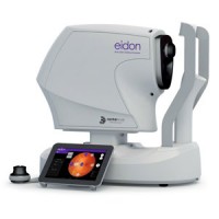
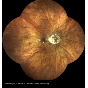
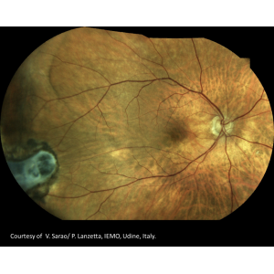
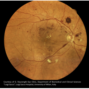
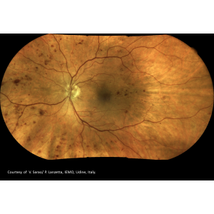
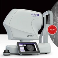
EIDON is the first system to combine the advantages of SLO with the fidelity of true color imaging, setting new performance standards in retinal imaging. EIDON provides unsurpassed image quality, 60° field in a single exposure, a unique, live, confocal view of the retina, three different imaging modalities and dilation-free operation, all integrated in a versatile system that provides new opportunities in retinal diagnostics.
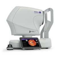
The device is operated via a tablet with a multi-touch, high resolution, color display; it works with a dedicated software application and operates as a standalone unit. A joystick is provided when manual operation of the device is desired.
SLO systems are superior to conventional fundus cameras in many ways, as they exploit a confocal imaging principle which limits the effect of backscattered light from deeper layers and provides enhanced image quality. Another major advantage of SLO systems is that they operate with much smaller pupils than conventional fundus cameras.
Eidon, being a confocal optical system, is able to perform high quality retinal images: increased sharpness, better optical resolution and greater contrast when compared to traditional fundus camera imaging.
This technology captures retinal images of preserved quality even in cases of media opacity.
LED flash technology guarantees maximum patient comfort as it uses a low power light source.
SLO systems are superior to conventional fundus cameras in many ways, as they exploit a confocal imaging principle which limits the effect of backscattered light from deeper layers and provides enhanced image quality. Another major advantage of SLO systems is that they operate with much smaller pupils than conventional fundus cameras.
Eidon, being a confocal optical system, is able to perform high quality retinal images: increased sharpness, better optical resolution and greater contrast when compared to traditional fundus camera imaging.
This technology captures retinal images of preserved quality even in cases of media opacity.
This in turn reduces pupil constriction and facilitates the test on non-cooperative subjects.
Key Features
Multiple imaging techniques with different light sources: white LED (440-650 nm), near infrared LED (825-870 nm)
Automated Stereo Imaging of the Optic Nerve Head
60° retinal pictures in a single exposure, wide field Mosaic up to 110° in automatic mode and 150° in manual mode
Manual Cup-to-disc calculation
Extreme ease of use: patient auto-sensing, auto-alignment, auto-focus
All-in-one compact design, no additional PC required
Simple Networking Options, for both remote data review and data backup
Benefits
True color, Red Free and infrared confocal images
Super-high resolution and contrast
Capability to image through cataract and media opacities
Dilation-free operation (minimum pupil 2.5 mm)
Wide Field imaging (60° in single exposure and up to 150° with Mosaic function)
Optimal exposure of the optic disc
Exam time less than 1’ per eye (single field)
From Fully automated to Fully manual mode
User friendly software interface
| Image acquisition | |
| Non-mydriatic (minimum pupil size 2,5 mm) | |
| Field of individual image | 60° (H) x 55° (V) captured in a single exposure [Center of Eye Angle of 90° (H) x 80° (V)] |
| Sensor resolution | 14 Mpixel (4608 x 3288) |
| Light source | infrared (825 - 870 nm) and white LED (440 - 650 nm) |
| Wide field Mosaic | out to 110° (H) x 95° (V) in automatic mode [Center of Eye Angle of 160° (H) x 135° (V)] |
| Wide field Mosaic | out to 150° in manual mode [Center of Eye Angle of 210°] |
| Working distance | 28 mm |
| Resolution | 60 pixel/deg |
| Optical resolution on the retina | 15 microns |
| Pixel pitch | 4.9 micron |
| Imaging modalities | color, infrared, red-free |
| Dimensions | |
| Unit Size | 360 (W) x 590 (H) x 620 (D) mm |
| Unit Weight | 25 kg |
| Power supply | |
| 100-240 VAC, 50-60 Hz | |
| Power consumption | 80 W (see label) |
| Accessories | |
| External power supply | |
| 3D Joystick with holder | |
| Tablet with holder and USB cable | |
| User manual | |
| Lens cap | |
| Removable forehead-rest | |
| External fixation | |
| Other Features | |
| Imaging modalities | color, IR, red-free |
| Automatic operation | auto-alignment, auto-focus, auto-exposure, auto-capture |
| Auto-focusing adjustment range | -12D to +15D |
| Dynamic, programmable internal fixation target, in every position of the field | |
| Tablet operated, with 10.1” multi-touch, color display | |
| Wi-Fi connectivity through tablet | |
| Ethernet connection through device | |
| Patient presence sensor | |
| Hard disk | SSD, 256 GB |

