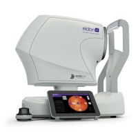
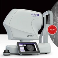
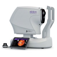
EIDON AF
Eidon AF represents the natural evolution of Eidon TrueColor Confocal Scanner, including all its features and functionalities, preserving its unsurpassed quality of image, and adding autofluorescence imaging capabilities.
Eidon AF with Autofluorescence capability is now an extraordinary tool for obtaining multiple types of highvalue information from multiple imaging modalities:
White illumination is able to provide high-quality TrueColor imaging
Red-free is useful to enhance the detail of the retinal vasculature and retinal nerve fiber layer
Infrared light provides information corresponding to the choroid
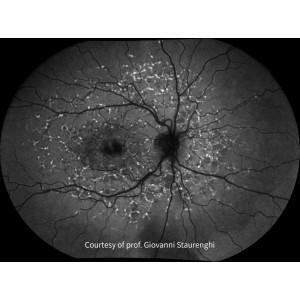
Autofluorescence allows the assessment of the Retinal Pigment Epithelial (RPE) layer
The importance of Autofluorescence Imaging in Clinical Practice
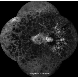
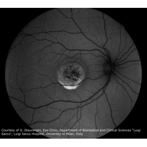
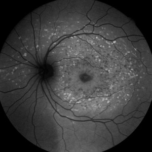
Fundus Autofluorescence (FAF) imaging is a non-invasive technique which provides information on retinal metabolism and health, through showing changes in the integrity of the Retinal Pigment Epithelial (RPE) layer. FAF imaging may help to understand metabolic alterations of the RPE in the pathogenesis of several retinal disorders. FAF provides these clinically useful information by taking an image based on the distribution pattern of fluorescent pigments accumulating in the RPE, such as lipofuscin. RPE dysfunction is visualized as an increased FAF signal, that is bright areas in the image, corresponding to lipofuscin accumulation RPE or photoreceptor death is visualized as a decreased FAF signal. That is dark areas in the image, corresponding to lipofuscin absence
Features
Includes all features and functionalities of EIDON technology, adding auto-fluorescence imaging capabilities
Provides confocality based on scanning line
Presents the highest pixel resolution (14 MP sensor) for a confocal system
Captures 60° autofluorescence image with a single flash of light
Mosaic function provides Wide field up to 110° autofluorescence images
Operates in fully automatic or manual mode
Benefits
TrueColor, Autofluorescence, Infrared and Red-free Confocal images
High details and contrast in the same device
High fidelity of the image without image averaging
Short exam time and enhanced patient confort
Provides a panoramic view of the retinal autofluorescence (up to 110°!)
Easy to use, allows to speed up patient workflow.
| Image acquisition | |
| Non-mydriatic (minimum pupil size 2.5 mm) | |
| Field of individual image | 60° (H) x 55° (V) captured in a single exposure |
| Sensor resolution | 14 Mpixel (4608x3288) |
| Light source | near infrared (825-870 nm), blue (440-475 nm) and white (440-650 nm) |
| Working distance | 28 mm |
| Resolution | 60 pixels/deg |
| Optical resolution on the retina | 15 microns |
| Pixel pitch | 4.9 micron |
| Other Features | |
| Imaging modalities | color, IR, red-free, autofluorescence |
| Automatic operation | auto-alignment, auto-focus, auto-exposure, auto-capture |
| Auto-focusing adjustment range | -12D to +15D |
| Dynamic, programmable internal fixation target | |
| Tablet operated, with multi-touch, color display | |
| Ethernet connection through device | |
| Hard disk | SSD, 256 GB |
| Dimensions | |
| Weight | 25 Kg |
| Size | 620 X 590 X 360 mm |
| Power supply | |
| 100-240 VAC, 50-60 Hz | |
| Power consumption | 80 W |

